Tag Archives: biology

CGM overestimates glucose in comparison to capillary sampling

I have heard that continuous glucose monitors (CGM) are becoming popular among individuals interested in their responses to diet, and their health risks.

In relation, this appears important:

A pre-proof from the American Journal of Clinical Nutrition

https://ajcn.nutrition.org/article/S0002-9165(25)00092-9/fulltext

Note, only one brand and model of CGM was used in the study. Significant variations in CGM values among different individuals tested under similar conditions were also observed. Some caution in generalizing findings is justified.

Thebgroup reports a mean overestimation by CGM versus capillary sampling as 0.9 mm/l, which is 16.2 mg/dl.

And this contributed to larger CGM calculated values for area, in area-under-the-curve calculations; e.g. the duration of hyperglycemia after a glucose challenge or a meal.

Conversion of mmol/ L into mg/dL is x18. Clear circles are CGM values, red are those values adjusted for difference relative to capillary samples.

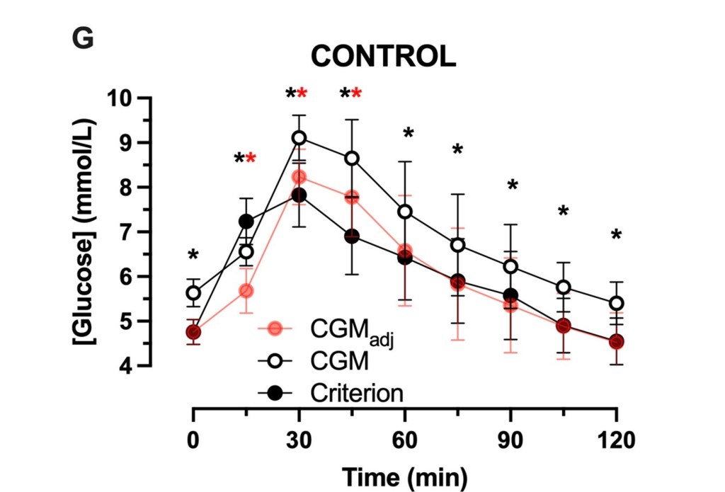
“CGM-estimated fasting and postprandial glucose concentrations were(mean±SD) 0.9±0.6 and 0.9±0.5 mmol/L higher than capillary estimates, respectively(both, p<0.001).”

“The increase in glucose concentrations measured with CGM versus the criterion method resulted in a >3.8-fold increase in the time spent above 7.8 mmol/L…”

“CGM overestimated glycemic responses in numerous contexts. At times this can mischaracterize the GI [glycemic index]. In addition, there is inter-individual heterogeneity of the accuracy of CGM to estimate fasting glucose concentrations. Correction for this difference reduces, but does not eliminate, postprandial overestimate of glycemia by CGM. Caution should be applied when inferring absolute or relative glycemic responses to foods using CGM, and capillary sampling should be prioritised for accurate quantification of glycemic response.”

Mitochondria are Mobile

Saturday, February 22, 2025

Over generations or during a lifespan, whole and individual, or in fragments 

  • Early in life’s origins, as now, there were many different lineages of living cells with each bearing individual adaptations. Keeping alive requires resources to be brought into a cell; some cells evolved a capacity to engulf not only useful nutrients but also absorb other cells. A cell engulfed by another would likely be broken down for resources unless it had evolved effective defenses.
  •  At this microscopic scale, living matter may show unexpected persistence. Sometimes an engulfed cell could defend against breakdown and partially survive, continuing its life as a passenger, perhaps a parasite, or better, a valuable symbiont. Evidence for this process as it happened then and continues now, is visible under a microscope.
  • When this survival by symbiosis merged cells with different but compatible abilities, a new form of cell could result, opening a trail for new kinds of life to appear and expand.
  • Our bodies, and much of the life we see around us, are built of cells that were originally such a new lineage, one bearing a fortunate combination of abilities, with its living components evolving adaptations to each other. That cooperation is ongoing then and now, with the symbiotic partners mutually changing over time.
  • Mitochondria became a symbiotic partner in cells in this way. They were once a type of bacterial life which had evolved a highly efficient method of producing useful energy from nutrients. Cells engulfing mitochondria that could keep this new passenger’s energetic function harnessed and regulated for mutual purposes benefited tremendously. The surplus energy acquired relaxed former limits on growth and genetic information capacity in the new lineage of cells.

Cell Nucleus, Mitochondrial Nucleoid

  • The dual sets of DNA in this new lineage became mutually adapted over time. The larger genetic set, the cell nucleus, was protected by a membrane and packaged with specialized protective proteins, while most of the original mitochondrial gene set was copied over into the nucleus during this long process of adaptation. The DNA remaining behind within the mitochondria reflects its bacterial origin: it is circular, smaller, and lacks a protective membrane. There are usually several copies of this remnant genome within each mitochondrion, and many mitochondria within each cell.
  • As a result of this DNA transfer, the organelles can no longer independently replicate a full DNA set to reproduce. Instead, their reproduction is organized as a cooperative readout of nuclear and mitochondrial genes, leading to assembly of new mitochondria. The process is called mitochondrial biogenesis.
  • An individual mitochondrion seen at a given moment will appear small as compared with the cell nucleus. They are a challenge to observe in a living cell. But when observed closely in vivo, movement and change are ongoing. At their molecular scale they host multiple sets of modular protein components that can contain and catalyze a myriad of biochemical reactions, including capturing potential energy from nutrient breakdown for use throughout their host cell. At the same time on the microscopic scale, they are in movement, with individual mitochondria fusing their membrane structures to each other in groups, forming large networks before separating into individuals again. 
  • In this ongoing fusion and fission process, a mitochondrial network’s contents, including the many sets of DNA and energy production machinery can be shared, mixed, and then redivided. These components are constantly active and are subject to much wear and tear from energetic chemical reactions and their byproducts. Like a handful of coins pulled from a pocket, some components are more battered than others, and consequently less functional.
  • When a network separates again into its parts, the reshaped mitochondria are not the same as before and are not identical or equal to one another in function as a result of the shuffling of the active components they carry. Most may average out in their function if they contain enough intact components, but some will have received more worn and damaged parts.
  • Also ongoing, there are cell operations that can find and tag defective organelles, worn and misfolded proteins, and other waste products, bagging these up for disposal in a process called autophagy. It appears that the less functional individual mitochondria are more often tagged for disposal. Careful observations of these processes in action together suggest an evolving quality control mechanism acting on a cell’s contents and organelles, through waste disposal coping with wear and malfunction and so sustaining cell viability.